Tuesday, February 28, 2006

Angioplasty: The Malaysian Story

As we grow older, we tend to sit in the corner, and remember how life has been and had been. Some of us get an urge to record what happened for posterity.

Upon graduation from the University of Malaya in 1975, we were at the stage of staying congenital heart disease with the use of serial "park" films at University Hospital Kuala Lumpur (UHKL). The X-ray filming equipment was donated to us by the Americans and they sent Dr J.W. Kennedy (of LV cineangiogram LV function calculation fame), over to teach us how to do diagnostic studies on congenital heart disease. Dr J.W. Kennedy was very instrumental in developing cardiology in Malaysia. Besides me, Prof H.O. Wong, Dr K.T. Singham and Dr A. Masduki all benefitted from his training.

Our emphasis then was congenital and valvular heart disease, as these disease were then highly prevalent in Malaysia. It was closely linked to out pioneering cardiac surgery program in UHKL (that's another story). Coronary artery disease then was not a priority and no "coros" were done.

I left UHKL for Glasgow in 1977 to write my MRCP-UK. The present day trend of being offered a job by the host nation was prevalent then too. Upon completion an offer for a cardiology job in Glasgow Royal Infirmary was taken up and I trained under Dr Ross Lorimer and Dr Ian Hutton, in coronary arteriography.

At the same time Dr Nik Zainal was training in St Mary's Hospital to do coronary angiogram. While we were angiographing the Judkin's way they were , at St Mary's, doing "Coros " the Sone's way. He returned to HKL in 1980 to start the coronary program in HKL, and I rejoined UHKL the same year. At that time, Dr KT Singham was trying to start a coronary program, but the UHKL program was not as smooth as HKL, partly because of cardio-surgical facilities and interest and also personnel problems in UHKL.

There was no co-ordination between the HKL program and UHKL cardiology consultants. The early 1980's were also the dawn of angioplasty. Dr Gruentzig had done the first angioplasty in 1977 in Zurich, Switzerland, and Dr Richard Myler and Dr Simon Stertzer were also doing angioplasty in California. Some of our wealthier patients were being referred to the USA for angioplasty accompanied by some of our refering physicians on a fully paid basis when they flew with the patients.

Most of the patients then were referred to Dr Myler and Dr Stertzer in San Francisco Heart Center, in Seton City. Dr Sterzer would occasionally visit South East Asia to see some of his patients. On one of this visit to KL in 1982, he performed an angioplasty in UHKL. That was probably the very first angioplasty done in Malaysia. There was no attempt at a program but just the once a year case on the basis of the patient being a VIP who could pay. This went on for a while.

The equipment in UHKL was old and the film speed was hardly adequate for good quality coronary angiograms. There was no plan to start interventional cardiology although it was clearly gaining popularity in America and Europe. In 1983, Dr Gruenzig himself visited KL to give a talk to a packed audience. This was to be his last trip to KL as he met an untimely death in 1985 following an airplane crash. What a loss to the interventional world.

I struggled to get a coronary program started in UHKL without success. The obstruction was phenomenal. At that time the game in town was the occassional visitor swooping in and to do the occasional case, usually for VIPs, and that was it. This was an unthinkable way of doing things. All attempts were met with the response that medical lecturers should only teach and go on general medical calls. Ironically enough it was 1984 when I chose leave this Orwellian nightmare to join private practice. What a symbolic year.

In the interim I had been tracking the progress of angioplasty. There were training programs held regularly in the US. Most importantly, there were included "live cases", so that we could see and learn, through live transmission. That was 1988. The courses were well conducted and the registry data showed good success rates, with good symptom relief.

There was no reason Malaysia should be left out of the benefits of having an angioplasty program. In Jan 1988, I pulled together a small team, consisiting of a trained cardiologist from HKL (Ministry of Health), one from UKM (ministry of education) and myself (private practice), to go to San Francisco to attend a live demo course in April 1988, and then decide what was the best thing to do to bring the technique back to Malaysia.
On Comments and Questions

This is just a quick administrative detail. Hopefully readers who leave comments or questions can leave an email address handy for us to reply to. Alternatively if you are concerned about leaving your email address, please do not hesitate to email us and we will post a reply here with a link to your email. This will help us ensure everyone gets a reply. Thanks for understanding.

Grand Rounds 28

Thanks to the folks at Grand rounds for featuring one of our posts in Grand Rounds 28.

Rising cost of cardiac healthcare

The latest issue of the European Heart Journal carried a study by the Health Economic unit of Oxford University on the cost of cardiac care in Europe in general and UK in particular. In 2003, the European Union spend 169 billion Euros on cardiac care (about about 230 euros for every man, woman or child). This is alot. Cardiovascular disease and stroke accounted for two thirds of the Cardiovascular death and 47% of the health budget.

CVD (Cardiovascuar disease) acounted for 268.5 million working days loss with 1:100 european is hamper by CVD. 17% of UK's healthcare budget is spend on CVD.

These are very telling numbers. Unfortunately we do not have our own numbers, but we can make a few succinct observations.

Obviously, people wish to live longer, whether it is Europe, UK or Malaysia. The public must know that to live longer and better costs money. The government may not be able to pay for everything all the time. We all know that no government who hopes to be democratically elected will ever say such things. The cardiac cost to the national budget is just too high, if you want the best, the "rakyat" may have to pay something. The Malaysian National Health Insurance Scheme is so long in coming, that one wonders whether it will come during our life-time.

That the best and most cost efficient thing to do is for the government to go full steam ahead with cardiac preventive programs. We could start with a no added salt diet campaign. There could also be a label all junk food campaign, so that the calorie, fat and salt content is fully declared. Also there should be a low sugar campaign, for example reducing the amount of sugar in carbonated drinks, in an attempt to reduce obesity. Reducing the incidence of hypertension, obesity and diabetes, will reduce coronary artery disease. Of course, as with all legislation, enforcement is important, so that campaigns can bear fruit.

All males above 40 and all females above 50 should go for a cardiac checkup, including a stress ECG, paid by the company, by Socso, or by EPF. This strategy will allow us to detect cardiovascular disease early. Test early, test often. This way we catch it when treatment is easier and usually less costly.

Of course we all want to live longer and better. But to expect this to cost almost nothing is unrealistic. Prevention is better than cure, and much cheaper too.

Sunday, February 26, 2006

Calculate your chance of dying

The Journal of American Medical Association published, on the 15th of February 2005, a thought provoking article by Dr Sei Lee. He had conducted a survey in 1998 on 11,701 patients. Using the data, he then statistically modelled a persons risk of dying over four years, depending on 12 important risk factors. These 12 risk factors are given risk weightage. At the end of the risk assessment, the total number of points accumulated is then used to tell you the risk of dying over 4 years.

Let us take a look at the model (it is interesting). The 12 risk factors and their weightage are :

1. Age :
60-64 yrs - 1point
65-69yrs - 2 points
70-74 yrs - 3 points
75-79yrs - 4 points
80-84 yrs - 5 year
>85 yrs - 7 points.

2. Sex : Male - 2 point,

3. BMI :
Below 25 - 1 point

4. Diabetes: 2 points.

5. Cancer: 2 points.

6. Requires oxygen use at home: 2 points.

7. Congestive heart failure: 2 points.

8. Cigarette smoking in the past week: 2 points.

9. Difficulty bathing/showering because of a health or memory problem: 2 points.

10. Difficulty managing money, paying bills, keeping track of expenses because of a health or memory problem: 2 points.

11. Difficulty walking several blocks because of a health problem: 2 points.

12. Difficulty pushing or pulling large objects like a living room chair because of a health problem: 1 point.

0 to 5 points - Less than 4% risk
6-9 points - 15 %
10-13 points - 42%
greater than 14 - 64%

Well those are the details, if you wish to calculate your own risk. I score 2 points, so I should be OK in the next 4 years. But is this what we should learn from this fine piece of work? We could learn a few other lessons. We could learn that these are important factors that would affect our health and well being as we gray, and lose our hair. Take note of the risk factors and prevent some of them if possible, eg. cigarette smoking, lose weight, and diabetes. Maintain good physical fitness through regular exercise. Know the negative factors and prevent them and stay healthy. We also learn that we should do the mentioned everyday activity like walking a few blocks, and also note that mental health is as important as physical health. Maintaining good memory is as important as maintaining physical fitness. Basically, note the risk ahead and do your best to avoid them. All the best for your next 4 years, and all future 4 years.

Saturday, February 25, 2006

64 Slice MSCT - Friend or Foe?

This blog had an earlier post which covered the 64slice MSCT among other things. Interestingly enough, this was also covered by JAMA in its Feb 15th issue.

It is well known that we probably have the highest number of 64 Slice MSCT (64 MSCT) scans per 100,000 population in the world. For some reason, there are almost 6 such machines in the Klang valley. Undoubtedly, the 64MSCT does have good clinical use, as it does give better definition, thinner slices and requires less scan time. This in turn means less breath holding for the patient and less motion artifacts for the doctor. The 64MSCT also allows for 3D volumetric reconstruction. The software is much more sophisticated.

Balanced against these good points, there is the tremendous amount of radiation the 64MSCT exposes the patient to. Almost the equivalent of 500 plain CXRs per cardiac angiogram scan, and 2000 plain CXRs if you do a total body scan from head to toe. I wish this was told to the patient, in terms of full dislosure. In fact there is some concern that females and males with chest scans run the risk of Breast Cancer.

This is not the biggest problem. The MSCT has also brought about a new medical problem of "turf division". For a long time, Xrays and imaging techniques generally have been under the responsibility of the radiologist, who are experts in Xray shadows and their interpretation. They better understand the physics of Xrays. When you see an image, they are better trained to differentiate real from artifacts. Even the expert radiologist sometimes is uncertain and make mistakes, what about a cardiologist who visits a scan center for 2 weeks and become an "expert", reporting 64MSCT?

This logically leads to the next question of how good the scans are, in particular the non-invasive coronary angiogram. Well, in the best of centers, where dedicated radiologist/MSCT trained cardiologist working on these scans day in day out, the positive predictive value of about 93-95% (these numbers vary depending on the part of the coronary tree we are examining) and a negative predictive value of 97-98%.

What are our own findings, given that many of our scans are interpreted by consultants who report the 64MSCT part time. There is no data coming out, apart from the correlation work being done in Sarawak GH, of which results we are eagerly awaiting. The other big issue is that a medical diagnostc technique with known dangers are being allowed to be marketed to the unknowing public, without full disclosure of the risk. This the authorities must take note.
So the 64MSCT, friend or foe, you decide?

Friday, February 24, 2006

Which placebo produces more effect?

The pills lose out to devices in terms of placebo effect.

The 1st Feb. 2006 BMJ carried an interesting study comparing the placebo effects of a sham acupunture needle, to placebo pills. After the run in periods, where the placebo effects as in subjective pain scores were identical, over the longer 6 weeks of the trials, the sham device was clearer more "placebo-ic" then the pills. It was also interesting to note that the authors also concluded that whatever adverse events were discussed at the consent, were also reported more frequently at the follow up.

What does this mean. Well, it could be that it is easier to convince patients of placebo effects if you use a device, or do something physical, rather than using a pill. Doing something sells better (isn't this true). Also, if you want adverse events reported, suggest it during the consent stage. Isn't that true? Well now we have some evidence. It was a small study, 270 patients only, but otherwise, quite a well carried out study.

Heart Tests: Coronary angiogram - Non Invasive

Here is part 21 of the heart of the matter. Part 20 is here and the dislcaimer is here.

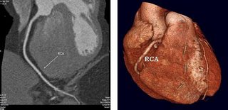
This is a picture of a 64 slice multi-slice CT coronary angiogram


Computerize tomography is a form of X-ray imaging of the heart. It involves taking serial X-rays of the heart, thinly slicing the heart using an X-ray knife, then re-constructing them (3D reconstruction) with the use of sophisticated computer software, to form a virtual heart. The pictures indeed look seductive.

Heart arteries can be targeted by localizing the arteries with contrast dye. We can then also re-construct the arteries. This sounds simple enough, except that it is not quite so simple. Is what you see, the true picture? Or is it not?

One of the biggest problem with sequential X-rays of the heart is motion. The heart moves, from about 70 beats per minute, to anywhere near 100beats per minute when the subject is excited, anticipating the CT scans. The increased heart rate will cause motion artifacts (the X-ray camera is filming slower then the target object, that is in motion).

The other technical problem, is localizing the heart arteries with the dye. Without the dye, the X-ray camera cannot differentiate the arteries. Since the dye is injected into an arm vein, it takes time to circulate to the heart artery, and in the process, also becomes diluted, so that the localizing of the heart arteries, will not be as precise as the selective coronary (invasive) angiogram.

The other obvious difficulty in heart artery imaging is the tortuous, twisting and winding course of the heart artery. No two persons have the same size artery, running at exactly the same course. More so when we know that Asian arteries are smaller then Caucasian arteries, and maybe more calcified.

Therefore, when the CT scanner takes pictures of the heart arteries at standard projections (guided by software meant for Caucasians), it may X-ray slice arteries at uneven places, giving false information. The arteries may contain speckles of calcium which cast interfering shadows called "calcium artifacts", or the heart arteries may be small, and cutting it unevenly, may cause impression of false blockages, or miss blockages that are there.

The patient is under the X-rays scanner for about 20-30secs. That's how long it takes for an average scan, and the rest of the data is acquired post-procedure, (off-line heart artery reconstruction). The reconstructions accuracy is heavily dependent on the adequacy of the initial on-line scan acquisition (poor initial scan acquisition means poor data input, and so false picture reconstruction. Garbage In Garbage Out is something they still teach in computer schools).

Suffice to say that there is a " professional learning curve " required, to try and learn the skills of proper scan acquisition for our Asian arteries, which are somewhat different, from the Dutch, German, or American arteries. We all want to see our arteries to make sure that they are okay, but be aware that you may see a picture of your heart arteries, but what you see may not exactly be the true picture of your arteries, given all the artifacts and limitations and assumptions made by the scanner software.

Basically, the multi-slice CT angiogram, is an evolving technique, and even now, refinements are being made. The very first scanners were single slice CT, mainly to assess calcium score. Then came the 4 slice CT, then the 8 slice CT, then the 8slice, then the 16 slice. The current scanners in Malaysia are mainly the 64 slice. The 128 slice scanners are already on display waiting to be marketed. I hear that a 256 slice prototype scanner by Toshiba will be exhibited soon.

The progress in technology is amazing. The high radiation exposures are being looked at critically. Suffice to say that the early machines exposes the subjects to (for a CT coronary angiogram) the equivalent of 500 chest X-rays. A total body CT scan, exposes the subject to almost 2,000 chest X-rays. Certainly not small amount of radiation.

To slow the heart rate (to avoid motion artifacts), the subjects are given beta-blockers. Some subjects can't tolerate the beta-blockers and fatalities have been recorded.

Calcium artifacts are difficult to overcome.

Poor on-line technique cause some scans to be inadequate for interpretation, or some sections of the heart artery to be inadequate for interpretation. In fact, some medical studies show that as much as 5-10% of heart artery segments may not be assessable using this scan technique.

Perhaps one of the biggest issue with this CT angiogram, is the lack of medical data, what with these new machines coming out so fast that it's almost tempting to belief that such upgrades are driven by sales and marketing, not so much by medical need. "Evidence based medicine" type doctors find it difficult to recommend the use of this machine for general population screening because of a lack of medical evidence of it's usefulness and it's dangers We believe that it does have a role to play in the assessment of CAD but surely not population screening of healthy adults.

Although invasive coronary angiogram is invasive, but with advances in technology, it too has become minimally invasive but remains definitive and is still the "gold standard" for the diagnosis of CAD.

To summarize

  1. The invasive technique is invasive as it's name implies. MSCT is non invasive.
  2. The invasive technique has a 100% accuracy for both positive and negative prediction purposes. The best MSCT shops are scoring around 92%-95% (postive) and 95% (negative) in terms of predictive value.
  3. The radiation risk of invasive angiograms are less and the exposure risk for MSCT can only increase.
  4. Invasive techniques avoid motion and calcium artifacts in the image. MSCT carries these artifacts. Worse yet, it may not be obvious since the image is post-procedure processed by software not customised for your ethnic group.
  5. The cost of an invasive angiogram is more at approx RM 4000 (USD 1000) with the cost of MSCT is at around RM 2700 (USD USD 700).
  6. The invasive equipment you use is probably mature and any changes would be minor. The MSCT equipment keeps on changing at an alarming rate.

Heart Tests: Coronary angiogram - Invasive

Here's part twenty of our series entitled the heart of the matter. Part 19 is here and the disclaimer is here.

This was accidentally discovered by Sones in 1965 at the Cleveland clinic angiogram laboratory, when he accidentally injected dye into the right coronary artery, got some nice pictures, and the patient survived. You must understand that before 1965, it was considered anathema to instrumentate the coronary artery as it was thought to be fatal. The heart was sacred.
With Sones showing us the way in 1965, we have now developed a whole new field of cardiology called interventional cardiology. We boldly intervene inside the coronary arteries, saving many lives (more about that later).

Heart arteries are very small, 2-3 mm in diameter, and so the common X-rays cannot see it. Therefore to visualize the heart arteries, we need to pass a small rubber tube (catheter) into any of the major arteries eg femoral artery or radial artery, of the body (they all lead to the heart ultimately), and with the X-ray screening to guide us, move the rubber tubing (catheter) carefully into the opening of each of the major heart artery, magnify the artery, inject dye (contrast material) to outline the artery, and take pictures. As the arteries twist and turn unpredictably, it is often necessary to change our angles to try and film the artery in the right profile. Although there are standard views to take, we need to individualise for the patient.

We learn that no two persons artery flow exactly the same course. Some are longer, some shorter, some turn acutely, some turn gently, some have big branches, some smaller branches, some arise nearer the opening, some further, etc. All these variations must be adjusted for having seen the preceding shots. At the end of the picture taking, all pictures are stored in a CD (previously 35mm cine-films). The coronary angiogram usually takes a half hour, in experienced hands. The information obtained is invaluable.

The coronary angiogram is invasive, as it requires us to puncture an artery, and so requires the subject to be hospitalized for 6 hrs at least, if not overnight. There are potential problems with dye allergy and bleeding. There are also potential complications of heart attacks and strokes. There is a small (0.1%) risk of death.

Therefore, we do not advice coronary angiogram simply but only for those who have signs and symptoms of CAD or a high likelihood of CAD. We feel that if the patient has a disease (CAD), the 0.1% risk of death is worth taking, in the hope of helping him avoid a higher risk of death from CAD. With coronary angiogram, there is also the question of radiation. It is an X-ray technique. The radiation is about 500 chest X-rays. In a patient with CAD, the risk/benefit ratio is heavily in favour of the angiogram. If the patient does not have CAD (just for screening), then the risk/benefit ratio is not in favour of an angiogram.


This is an example of a coronary angiogram showing stenosis in the right coronary artery.

It must also be said that there are definite benefits from a coronary angiogram. You do make a definite diagnosis (coronary angiogram is the gold standard). It allows us to decide what is the best treatment strategy, and if angioplasty is the best strategy, it can also (if the patient and doctor wishes), be performed at the same seating. Diagnosis and management together.

Part 21 discussing non invasuve angiograms is here.

Heart Tests: Coronary angiogram

Here is part 19 of the heart of the matter. Please read the disclaimer here and you can find part 18 here.

Now is a good time to review the basics. Coronary: as for the heart, angios: blood vessels, gram: picture. A coronary angiogram then is to make a picture of the arteries of the heart.

In the invasive form we invade the body, as in cutting or passing tubes into the body. Just a simple injection into a vein is not generally considered to be invasive. There is almost no controversy about the position of the invasive coronary angiogram as the "gold standard" to diagnose CAD. As for the use or as some would allege, misuse, of the non-invasive coronary angiogram, it is certainly very controversial at the moment.

Much has been written about it recently, on the one hand by heads of professional societies and on the other hand by professionals who have equity stakes in the machines!!! It is not the purpose of this blog to join in the debate however much joy it may give the readers but hopefully I can provide a simple, balanced view on this subject and leave the readers to draw their own conclusions.

DISCLAIMER: I am not a stakeholder in any way in any of the 4 MSCT (multi-slice computerized tomography) machines around Kuala Lumpur, or the 8 machines around the country. The number of machines may have increased by the time you read this.

It is true that if you suspect that you may have coronary artery disease (CAD), no amount of blood tests or ECG or echocardiogram will convince you one way or the other, until you see your arteries. For a long time, before the advent of computers and their sophisticated software, we relied on the invasive coronary angiogram as the gold standard.

Presently, with our advanced computers, hardware and software, we can image coronary arteries non-invasively. The pictures of your heart and heart arteries are really seductive, but is what you see the real arteries or is it just computer generated virtual arteries brought to you via the miracle of software processing? What is the clinical correlation through clinical studies between the "gold standard coronary angiogram", and the CT coronary angiogram? Are there enough clinical studies? What is the international experience?

In part 20 we talk about invasive angiograms.

Thursday, February 23, 2006

Heart Tests: Radionuclide scan


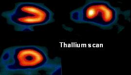
This is a test which uses radiochemicals like thallium and technetium to locate areas of deficient blood flow in the heart. The subject does a treadmill stress. At the peak of the exercise, the radiochemical is injected into his vein and a special nuclear imaging camera is used to image the heart at different angles, to check the adequacy of blood flow. Areas of inadequate blood flow will show as areas of low radioactivity (cold areas), detected by the camera. Knowing the blood flow pattern in the heart, we can co-relate the areas of "cold areas" with the territory supplied by the respective arteries. In those subjects who cannot exercise, we can use drugs through IV injections, to induce regional lack of blood supply.

It is a very popular test for the diagnosis of CAD in the developed countries, where nuclear reactors are always nearby. Technetium has a longer half life and so can be transported and stored for longer. Thallium on the other hand has a short half life (72hrs) and cannot be stored for long. This does give logistics problems in trying to arrange the tests. For both agents, there is the additional problem of "radioactivity" stigma and also radioactive waste disposal. That may be why these tests are only available in few medical institutions, and therefore not so popular.

However, in theory, it is a very useful test as it does have predictive accuracy in the 90% range, in good centers, and it is relatively cheap. We use it sometimes to help us sort out those patients with an equivocal stress ECG, who also has some coronary risk factors. We call this group patients with intermediate risk factors and equivocal stress ECG result. The radio-nuclide scans are also very useful in those who cannot exercise.

For those who are following our series, the heart of the matter, this is part eighteen. Part 17 is here and our disclaimer is here. In the next part of the series we discuss the coronary angiogram.

Wednesday, February 22, 2006

Heart Tests: Stress Electrocardiography

This is part seventeen of the heart of the matter. It's an ongoing series which we hope readers find educational. In part 16 we covered the echocardiogram and it can be found here. Our disclaimer can be found here.

A stress ECG is probably the most popular and frequently used test to diagnose heart artery narrowing. It is reliable and reproducible, having stood the test of time. It's physiological principles are easily understood giving it a very wide appeal. It is relatively cheap and yet has a predictive accuracy of about 70-80%, when done for subjects at risk of coronary artery disease. The more coronary risk factors the subject has, the more accurate the test, reaching almost 90-95% in patients who has anginal type chest pains.

Since this test is time tested, the machines are cheaper and the test is widely available. It has almost no risk if the subjects are asymptomatic and the resting ECG normal. The test results are easily understood by many practitioners and there is almost no mystery to the test.

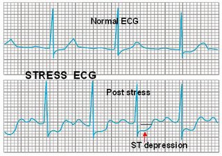
The stress test basically uses the ECG as an indicator of sufficiency of heart muscle blood flow. We have learned and validated the fact that insufficiency of heart muscle blood flow shows up in the ECG as ST segment change. The ST segment in an ECG is an indicator of heart muscle blood flow. A normal ST segment, signifies adequate blood flow, and a depressed ST segment, signifies inadequate blood flow. When there is blockage in a heart artery, the resting blood flow is sufficient for the resting heart, so the ST segment is normal. During stress, there is demand for more blood flow. Because of the narrowing, the supply of blood cannot meet the heart muscle demands, and the ST segment gets depressed. Simple and very straight forward.

However, if the area supplied by the narrowed artery is relatively small, the ECG detectors can miss. False positives and false negatives are occur with stress ECG test. But overall, for a cheap, clinically reliable, very low-risk cardiac test, with good predictive value (especially in the population at risk) the stress ECG is probably the best.

This picture illustrates how a normal ECG doesn't tell the whole story and the stress ECG can reveal more hidden facts.

In the next part we look at radionuclide scans.

Technorati Tags: , , ,

Tuesday, February 21, 2006

How good are placebo devices

The pills lose out to devices in terms of placebo effect.

The 1st Feb. 2006 BMJ carried an interesting study comparing the placebo effects of a sham acupunture needle, to placebo pills. After the run in periods, where the placebo effects as in subjective pain scores were identical, over the longer 6 weeks of the trials, the sham device was clearer more "placebo-ic" then the pills. It was also interesting to note that the authors also concluded that whatever adverse events were discussed at the consent, were also reported more frequently at the follow up.

What does this mean. Well, it could be that it is easier to convince patients of placebo effects if you use a device, or do something physical, rather than using a pill. Doing something sells better (isn't this true). Also, if you want adverse events reported, suggest it during the consent stage. Isn't that true? Well now we have some evidence. It was a small study, 270 patients only, but otherwise, quite a well carried out study.

Technorati Tags: ,

Heart Tests: Echocardiogram

This is part 16 of our continuing series, the heart of the matter. The last part is here and our disclaimer is here.

The echocardiogram is often use as a test for looking at the heart as a whole. It is very useful to detect structural abnormalities of the heart eg "hole-in-the-heart, valvular defects, crisscross hearts, blood clots in heart, etc. It is also good at diagnosing failure of the heart as a pump. It remains the investigation of choice for detecting congenital heart disease, valvular heart disease, heart muscle disease, heart lining disease, and also heart function.

Here's a picture of a color Doppler echocardiogram which we could use to diagnose structural heart disease.


However, there are a few things that it cannot do, eg. it cannot see heart arteries reliably, it cannot detect heart muscle lack of blood supply reliably unless coupled with provocation, and it cannot detect viability of the heart muscle reliably. In short, it is not so reliable in diagnosing early coronary artery disease. As we will mention later, stress echocardiogram (echo heart with provocation) can be used to detect CAD.

In the next part of the series we cover the stress ECG.

Sunday, February 19, 2006

Fish oil

[The doctor neither sells fish oil nor has an interest in fish oil companies]

One of the things I was taught in Preventive Health class was that Eskimos, although mainly carnivorous, have a very low incidence of CAD, just as Japanese in Japan. However, the Japanese in California have incidence of CAD that is almost similiar to white Americans. The Japanese in Hawaii have an incidence of CAD almost in between that of Japanese in Japan and Japanese in California. This important epidermiological observation gave rise to the principle that it must be the fish diet that Eskimos and Japanese have in common.

We now know that fish oils contain n3-polyunsaturated fatty acid (n3-PUFA), the important fish oil component giving the cardiovascular benefit. What is lacking is a large randomised control trial giving us conclusive evidence of this. Obviously these trials will never be as no corporation will spend the large amount of money to do this, as fish oil is non-patentable, so they cannot recover their expenditure.

There are smaller studies which conclude that in addition to primary prevention of CAD in Eskimos and Japanese, fish oil has also reduces the incidence of cardiac deaths, especially sudden cardiac death, following heart attacks. The risk of sudden cardiac death may be reduce as much as 45%. Besides that, fish oil also lowers plasma triglycerides and reduces the coagubility of blood, probably by lowering serum fibrinogen. There is some evidence that the n3-PUFA is incorporated into an atherogenic plaque, stabilising the plaque. Maybe that is why fish oils have been shown to lessen angina pectoris.

If you wih to take fish oil tablets, please know that you may have to take 1-2 grams daily, or take enough to smell fishy. Always be aware that there is increasing concern about the dangers of heavy metal contamination in fish oil. For those who would like to take fish, please note that different fishes have different concerntration of n3-PUFA. The deeper the sea, the colder the sea, the greater concentration of n3-PUFA in the fishes.

Basically, fish oils are good for CAD and sudden cardiac death prevention.. This is one nuticeutical agent with well proven cardiovascular benefits.

Thursday, February 16, 2006

Happy Valentines Day

Valentines day has come and gone. Much money must have been spent, buying gifts, chocolates being a favourite, and of course the romantic dinner with wine. It is indeed heartening to note that we all can enjoy a good bar of chocolate, but always prefer dark chocolates with a healthy amount of cocoa ( more then 70% ), if possible, because dark chocolate contain a healthy dose of flavanols, that have anti-oxidant properties and so help the heart, and blood vessels. It may protect against cholesterol clogging the heart arteries and may in fact increase coronary blood flow and also lower blood pressure. If you like nuts (referring to food and not choice of partner), choose walnuts, almonds and macadamias, all of which contain healthy plant fats, including plant omega-3 fatty acids.

As for dinner, Japanese is great for the heart, or fine dining Italian or Mediterranean with a good glass of wine. The Japanese diet and Meditteranean diet are cardiac friendly. The role of red wine, in increasing HDL-cholesterol and protecting the heart has been well documented. Fresh deep sea fishes obviously have good amounts of omega 3 fatty acid. Some Japanese green tea as you await your food is also good for the heart.

Yet always be aware that accompanying the bar of dark, dark chocolate, and the nuts, and the white potato are alot of calories. The dark chocolate is good for the heart , but the extra 500 calories per bar of chocolate may take a while to trim off and may require extra hours in the gym. HAPPY VALENTINE's DAY.

Tuesday, February 14, 2006

Not all heart attacks are typical

We have written previously that 40% of heart attacks strike without warning and sudden collapse is the first and only manifestation. Apart from these sudden cardiac death or Primary Ventricular Fibrillation (1o V.Fib.), alomost 30% of in the male risk age group and almost 50% in the females risk age group, may have very atypical symptoms when presenting with a heart attack. This was highlighted in a piece of research done in Rotterdam, by the group in Eramus Thoraxcenter, and reported in the latest issue of the European Heart Journal. They studied about 4,000 subjects and made this interesting finding.

I remember my old professor of radiology who had suffered a heart attack while working. One morning, about 20years ago, we (the head of cardiology-UH, senior cardiologists and myself - a junior trainee then) were doing our angiogram list in UHKL. The head of Radiology, a 50year old sikh gentleman walked in and mentioned to the "boss" that he was not feeling well, and had some gastric upset. My "boss" sent one of us to get him some mist magnesium trisilicate. After our morning angio list we went to see the haed of radiology and he said that he was still not feeling well. We quickly asked him to lie down and did an ECG which showed an acute inferior myocardial infarct (heart attack). The point here is that even the chief of cardiology can have trouble diagnosing a heart attack on history taking alone.

The lesson is obvious, that if you are in the cardiac risk group viz, males >40years old, or females >50years old, who are also diabetes, hypertensives, obese, cigarette smokers, hyperlipidemias, and if you should feel off colour or generally unwell, do an ECG. There may or may not be chest pains. If after 6-8 hours and your are still off colour, do a second ECG. If the complaint is typical chest pains going down the inner aspect of the left arm, that's easy. If it is not so typical, have a high index of suspicion, then do an ECG. Yes, we are maybe over doing ECGs, but an ECG is so cheap and so harmless, yet can be so rewarding. There are certain CVS risk groups who are prone to atypical features of AMI, viz females, the elderly ( >70years ), the diabetics, those are recoverying from non-cardiac surgery, those who have co-morbidities like chronic asthmatics or chronic arthritics on steriods, etc. Whenever in doubt, see the nearest doctor, and always do an ECG, and if necessary, blood test like cardiac enzymes, and troponin T or I. Remember, in heart attacks, time is heart muscle loss.

Quizzify KUIS

Quizzify is apparently one of the top scoring Scrabble words and earns you up to 400+ points if played correctly. Now let's look at another KUIS.

Not so long ago (last week to be exact), we wrote about the falling standards of medical education, following an open debate in the mass media, between the Malaysian Director General of Health, Ismail Merican, and the Deputy Minister of Higher Education Fu Ah Kiow. Tony Pua , uber Malaysian education blogger, linked to it here and here (thanks for the links). The Director General was very concerned at the ballooning numbers of medical schools in the country and the falling medical standards.

Now we hear that the state of Selangor has decided to have a medical school as part of this latest institution of higher learning. It is amusing that the rector appointed, Datuk Haji Mohammed Isman, was most concerned about where to locate the teaching hospital. There was no mention about medical teaching staff, so vital to the success of a medical school.

They are still awaiting the MOH's development plan of new hospitals to see how to tie up with MOH hospitals as their training center. They are presently sharing a hospital at Kubang Kerian with Universiti Sains Malaysia.

The grand plan is to have a school of pharmacy (this makes sense), a nursing college (this also makes sense) and dental school. One really wonders where the teaching staff are coming from. I suppose they will be imported from external sources.

According to the press statement, this latest medical school will contribute towards the human capital development of our country. I wonder if rector Datuk Haji Isman is aware that we presently have 2,000 new doctors added into the country of 28 million, every year, and that there are presently 18 medical schools already. Has the Ministry of Higher Education ever considered that they could combine resources. By working together they may produce good quality doctors in reasonable quantities at lower cost.

When will we ever learn that we need a long term plan in higher education planning? This is not a numbers game and we need to produce doctors of sufficient quality for the nation. After all, isn't this a life a death matter?

I stand corrected when complaining about 18 medical schools. The number to complain about now is 19. I wonder how many more are awaiting annoucement.

Monday, February 13, 2006

Heart Check 1: See your doctor

This is part 15 of the series heart of the matter. Part 14 can be found here and the disclaimer can be found here.

The most important part of a heart check is the interview with the doctor. This must form the basis of all checkups. The present day factory style "go through a barrage of test" and then have someone interpret the results and prescribe health is medically incorrect. There is much that can be gained from having a doctor see you first and take a careful history and do a thorough physical examination, even if you feel well.

This simple clinic check will allow the doctor to make an assessment and arrive at a provisional diagnosis, know what aspect of the heart check is important, what to look for, prescribe test that are relevant and not do test that are irrelevant (be cost effective). For example, we often see 30-something females without significant CAD risk factors going for a stress ECG. This subject has such a low possibility of CAD that the stress ECG cannot be interpreted. We see respiratory function tests being done for subjects who cannot blow, stress ECG being done for hemiplegic who can hardly stand.

Moreover, some "screening test" carries risk which are not well highlighted to the subject. Radiation risk for example. Deaths has been known to occur with some "routine screening test". If you are a healthy adult without disease, going for a health check, any risk, no matter how small, from any test, be it radiation risk, medication risk from tablets taken before tests, physical injuries sustained while performing test that you cannot perform, etc. is unacceptable. Even one is too many.

After a clinic interview, as part of the checkup, the resting ECG and CXR usually give us invaluable clues about the heart. It helps the doctor to know what other test may be pertinent. These test (ECG and CXR) are simple and cheap, and gives good information. The CXR does have a small risk of radiation,(relevant to the pregnant female subject) but is virtually harmless otherwise. As you can see, some test do carry risks although small.

Blood tests too are important as they allow us to form a risk profile for the individual. Just as in insurance where we risk profile clients, so also in heart check-ups. The higher the risk profile, the more likely the presence of disease, and vice versa. Blood tests eg the lipid profile and the exclusion of diabetes and renal disease helps us to risk profile the subject. We can also mark out a person's risk for a heart attack by checking for certain high risk markers in the blood like hs-CRP. These blood tests are cost effective.

In the next part we discuss echocardiograms.

FIELD - Fenofibrate Intervention and event lowering in Diabetes

This was presented at AHA2005.

Diabetes mellitus has become a very important risk factor for cardiovascular disease. Diabetes seem to bring about more severe disease and also confers a worse outcome in any category of heart diseaase. It is for these reasons that diabetes has become a major risk factor in CVS disease and is essentially a CAD equivalent.

This is probably why, 5 years ago, Fournier (a French company) planned the FIELD study. They presented some findings at the AHA meeting in Nov 2005 and also published it in the Lancet (Dec 2005).

After following 9795 patients, half on standard therapy and half on fenofibrate and standard therapy for 5.5 years, they found no difference on the primary end-point of non-fatal MI (Myocardial Infarction) and CHD related death.

There was, however, a diference in the secondary endpoint of MACCE (Major Adverse Coronary and Cerebral Event), where there was an 11% reduction after 5.5 years, in the patients on Fenofibrate (lipanthyl).

What this all means is that taking lipanthyl with standard medical therapy in diabetics (including statins), does not lessen the risk of dying, but it certainly reduce the risk of non-fatal MI and also lessens the risk of re-vascularisation.

What is also important is that statins have a profund effect on the results. It is likely that the primary endpoint were not significant because many of the diabetics were already on statins as part of standard medical therapy, and that statins can be safely used in combination with fenofibrate. So patients with diabetics with elevated triglycerides, or even elevated LDL-C or low HDL-C, should be started on fenofibrate. It seems safe and prevents them from non-fatal MI and a need for re-vascularisation.

Sunday, February 12, 2006

Migraine and PFO

There is an strong association of strokes with migraine and often we see that, following a stroke, the migraine goes away. Some strokes are caused by paradoxical emboli that cross from the venous circulation to the arterial circulation by passing the lung filters, through a patent foramen ovale (PFO).

The PFO is present in all of us, but it uaually closes spontaneously, when we take our first breath. However, in about 20% of the normal, healthy population, the PFO may not close and can potential stay open. In fact, that seems to have been the case with Prime Minister Ariel Sharon of Israel, who initially suffered what appeared to be a TIA and a PFO was discovered.

His doctors anticoagulated him and decided to close the PFO using a percutaneous approach umbrella. However, he suffered a massive cerebral bleed before that could be done. The rest, as we say, is history. We certainly hope he recovers.

Back to our story that some patients with migraine had their their migraine cured with closure of the PFO. This is about to be tested in a large randomised control trial, ESCAPE, to be led by Dr Dodick of Scottsdale, and Dr Sommer of New York.

Can we cure migraine with closure of PFO? The ESCAPE trial will answer that. We await the results.

PFO is also covered here by Kevin MD.

Saturday, February 11, 2006

Door to needle time

The New Straits Times today carried an article on Kuala Lumpur Hospital's current strategy in the management of acute heart attacks. A casual glance makes one rather proud that the system has gained some award, albeit the Director-General of Health trophy. Humble as it is, it is still an achievement to win the National Quality Award 2005.

On closer examination however, I failed to see the "door to needle" time given to us. The door to needle time describes the time taken to administer the clot buster medication, eg IV streptokinase, or IV rTPA, after the patient has entered ER door. It is a test of the system's ability to respond to a heart attack emergency. The shorter the door to needle time, the more efficient the ER is to cope with a heart attack emergency.

We are all working towards a door to needle time of 20-30 mins. In an average first world country, the door to needle time is an average of 60mins. That is deemed too long and has resulted in certain countries training ambulance units and even firemen to fax an ECG from the home of the patient to the nearest hospital, following a 999 call, and upon confirmation by the cardiologist at the hospital, administer the clot-buster medication either at the home or in the ambulance on the way to hospital.

Obviously this is a serious attempt to shorten the door to needle time, because the sooner the patient suffering a heart attack receive the clot-buster, the sooner the artery blood clot go away, the sooner blood flow down the heart atery, the sooner the heart muscle recover. The has given rise to the term, "TIME IS HEART MUSCLE RECOVERY ".

Perhaps, the reason why Dr Jeyaindran, the critical care medicine specialist in KLH did not know the door to needle time was because he did not give his heart attack patients the life-saving IV clot buster. The newspaper write up stated that patients were given aspirin following their heart attack. Perhaps the newspaper reporter did not inquireif the patient then had to wait for a consultant to arrive and certify the use of the IV clot buster.

So while the door to aspirin time could have been short and commendable, the door to needle time is probably long. Aspirin, on it's own, without the use of the IV clot buster, opens the heart artery unreliably, and certainly is not standard cardiac therapy in an acute heart attack. What is even more important is that in the 21st century, a good tertiary cardiac care center would have a well equipped and well trained cardiac cath lab and team, to perform immediate angioplasty for the heart attacks.

Giving just sublingual aspirin alone, is poor therapy. Giving IV clot busters with aspirin is reasonable therapy (particularly in hospitals without a trained angioplasty team). The best therapy, especially an award winning therapy should be immediate angioplasty especially in a good tertiary care center.

Dr Jeyaindran and his team should be commended for their tremendous effort to improve the standards of care for heart attack patients. Beyond this achievement there is still a long way to go, how far this is we will leave you to judge for yourself.

Drug-eluting stents and angioplasty, the good, the not so goodand the really bad news

Undoubtedly, balloon angioplasty had revolutionised treatment of coronary artery disease (CAD). It has allowed us to treat symptomatic CAD easily without the need for GA and opening the chest. I am sure that Dr Andreas Gruntzig (the inventor of angioplasty) would have been very proud to see the number of patients he has helped with his technique of angioplasty.

The good thing about angioplasty is that we can control chest pains very effectively with minimally invasive teachniques. Of cause, plain old balloon angioplasty (POBA) has its problems, namely acute tear (dissection) in the treated artery, and also re-narrowing (restenosis).

The next logical evolution was the use of the bare metal, plain surgical grade stainless steel stent. This was better then POBA and acute artery dissection was a thing of the past. We no longer need a cardio-surgical team on standby. We now have "stent-by".

Bare metal stents (BMS) still had restenosis as a significant problem, running at about 15%. The next step came with the use of drug-eluting stents (DES). Whether this advance is an evolution or revolution depends on how you see the situation. There are two DES that are currently FDA approved, namely the Cypher stent (JnJ Cordis) and the Taxus stent (Boston Scienntific).

There are a few more waiting in the queue to be approved like the Endeavor stent (AVE Medtronic) and the XcentV stent (Guidant Corp). These DES has very low restenosis rates (<5%) even when we use long stents and multiple overlapping stents. We have been known to line the whole artery with 5-6 stents in the whole length of artery (full metal jacket) with good short and longterm results.

Except for certain subsets, angioplasty and stents have not been known to prolong life, but it does control chest pains well.

Now for the not so good news. Balloon angioplasty suffered from a 4-10% risk of acute artery dissection, causing acute heart attacks, which before the stent era, required emergency bypass graft surgery, which carried a 30% chance of dying and almost 100% chance of heart muscle damage.

While balloon angioplasty has a overall success rate of of about 95%, there are a group of CADs, the chronic total blockage, where the angioplasty success rate can be as low as 40-50% in some centers and is limited to 80-90% in the best of hands. Some blockages just cannot be angioplastied.

The nagging problem with POBA is restenosis. It was quite upsetting to see a patient where you performed a good POBA, where the acute angiogram picture was wonderful, and suddenly in 2-3 months, the artery blockage had returned, possibly worse then before.

This was ugly and usually upset the patient. Of course, with the BMS, the acute dissection and acute bypass problem was essentially solved. We no longer needed to send our angioplasty list to the Operation Room, and the cardiac surgical team could have a nice lunch.

However, the BMS brought a new problem of acute and subacute stent thrombosis. The stents are stainless steel and these attract blood clots, if the proper medical regimes are not followed. Procedural techniques also played a part. These happened on average about 1-4% of the time. This could be minimized by technique improvements and also the use of aspirin and ticlopidine as effective anti-thrombosis medications.

Then came the DES that we are still learning more and more about. With DES, the good news is that restenosis is now single digit (<5%). However, acute and subacute stent thrombosis still remain.

With BMS, the subacute stent thrombosis tend to come in the first 30 days. It tends to be more predictable. This enables us to try and figure out who the high risk group is. With the new DES, the subacute stent thrombosis could occur in the first 30days or as late as up to 1-2 years. Therefore the anti-thrombosis medications (aspirin and plavix/ticlopidine) had to be continued for much longer. This brought about the problem of non-cardiac surgery, should that be necessary on an elective or emergent basis following the use of DES. If these become necessary and the aspirin or plavix is stopped, there is an almost 10% chance of acute stent thrombosis. The DES are more costly and certainly more fuzzy stents.

This problem is so worrying to us, that if we know that the patient may need elective surgery after the angioplasty, we would prefer to use the BMS as a way to avoid the problem of acute stent thrombosis should the apirin or plavix had to be stopped for the elective surgery. With the use of DES, we also discovered a new problem of aspirin and plavix resistance (patients who may be taking the aspirin or plavix yet there is no clinical effect of these medication). With the widespread use of DES nowadays (in some centers, almost 95% of angioplasty includes the use of DES) we have learnt that there are cases of stent allergy, either allergy to the drug or the stent, or the polymer that the drug is bonded to. Stent allergy is still new and we are in the process of learning what we can.

While we improve by what is seemingly leaps and bounds, each and every step carries a high price. There is no such thing as a free lunch

And now for the bad news. As DES use becomes more widespread, more complex lesions are undertaken by the interventional cardiologist with greater and greater confidence as restenosis is low and indications for angioplasty become more liberalised. Cardiac surgery numbers drop worldwide. Is this better for our patients?

Thursday, February 09, 2006

Low fat diet shocker

The CNN headlines were surely encouraging to Mc Donald's and other peddlers of fatty foods. What a sensational headline "Low-Fat Diets Disappoint for Cancer and Heart Disease". Even the local newspapers picked up the call and published it. What joy to tweak the nose of the low-fat boosting killjoys.

Yet is that the whole story?

What's interesting about this article is that on casual review it seems to go against popular received wisdom. It has been often stated that low fat diets lessen heart disease and the study seems negate this assertion.

Read closer and it states that the study population was older than usual, and these were post-menopausal women. Perhaps we should call this a study of the effects of low fat diet on post-menopausal women.

It is food for thought that IN POST-MENOPAUSAL WOMEN, A LOW FAT DIET MAY NOT LOWER THE INCIDENCE OF HEART DISEASE .

This we need to note even if it doesn't make for sexy headlines.

Heart Checks

This is the fourteenth part of the heart of the matter. Part 13 is here and our disclaimer is here.

The saying is true, "prevention is better than cure ". Medicine has entered the era of preventive medicine, where we hope to create greater awareness among people of their health and teach them to know and appreciate good normal health, as opposed to sickness. In a way, we are trying to work ourselves out of a job. Incidentally. it is to this end that this column is initiated.

If all illnesses can be prevented and the population at large can follow all the preventive steps, then the need for doctors will be less. But alas, that will never be, although it remains our dream. Preventive medicine is also much more cost effective, and has lesser morbidity and mortality. It is our hope that this message gets through.

As a result of this paradigm shift, we see the setting up of many wellness centers whose aim is to check normal people and give them a clean bill of health. This is now a flourishing industry, spreading on to health tourism and mega-dollars.

Myths to correct

What are we looking for when we go for a heart check? Well, we are obviously trying to see if our heart is in good condition and if we are at risk from heart disease in the near future. Whenever I get a request for a heart check, I sense that the client (we can't call them patients as they have no illness), expects that after the heart check, he will be guaranteed that he is not going to suffer any cardiac events. This is the first myth that we must correct.

There is no one cardiac test (or even a series of test) that can completely exclude a cardiac event. Heart checks merely establish the state of your heart at that point in time with the current available tests, none of which have a 100% predictive accuracy.

The second myth to correct is that one test can tell all. That is simply not true. There are so many aspects of heart disease and so many kinds of heart disease, that it is not possible for one test to tell all. There is heart disease present from birth which we call congenital (eg hole-in-the-heart), heart disease from valve defects (valvular heart disease), heart muscle disease (cardiomyopathy), cholesterol heart artery disease (coronary artery disease), disease of the heart from hypertension and other related body maladies, and many others. Besides these, there are tests for the functional aspects of the heart, test for the structural defects of the heart, and test for the electrical aspects of the heart. No one test can tell all. How can they?

The third myth is that all tests are free of risk. Generally, the more sophisticated the test, the more likely the side-effects.

Most often, we are trying to establish three things.
1. Is the heart structurally normal
2. Is the heart functionally normal
3. In those at the CAD risk group, is there any evidence of heart artery blockage causing lack of blood supply?

In the next part we describe the first step in a heart check.

Case study on heart attack

This is a case study on heart attack and forms part thirteen of our series, the heart of the matter. Part twelve can be found here. Most of our readers should have already seen our disclaimer here.

Mr C is a 40 year old Chinese salesman, who works very hard and is under much stress to sell. He suffers from mild hypertension and is under treatment, though not very regular. One month ago, while jogging, he experience an unusual, tight band-like discomfort, over his chest. He began to sweat profusely, cold sweat. He felt dizzy. Realizing that something was not right, he called his colleague who was nearby, to come and help him. While he rested in the car, the colleague informed Mr C's wife, and drove him to the emergency room of a nearby medical center.

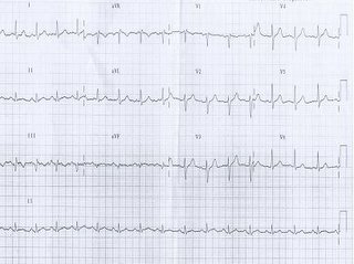
An ECG done there confirmed that he had indeed taken a heart attack. Mr C's ECG on arrival at emergency room, showing acute ST elevation suggestive of acute myocardial ischemia.


The cardiologist there decided to perform an immediate angioplasty (primary angioplasty). Mr C had total occlusion of his LAD artery. This was treated with a coronary stent. He had chest pains at about 10am. He reached the medical center at 11.30am, and the cardiologist opened the artery and implanted the stent at about 12.30noon (2hrs 30mins after the onset of chest pains). He was discharged about three days later showing full recovery of the heart muscle that was initially damaged. His ECG was virtually normal.


In the next part of the series we will be covering various checks.

Obesity and the heart

It is common knowledge that the Chinese like their food. In fact, the usual greetings when Hokkien Chinese meet is, "chiap pah ba?" (Literally "have you eaten"). To the Chinese a big tummy is a tummy of prosperity. Unfortunately, the potbelly is a danger to all of us.

As demonstrated again recently in subgroup analysis of the "INTERHEART" study and presented at the Nov. 2005 AHA annual scientific meeting. The original INTERHEART study (which involved 27,000 subjects in 52 countries) was first presented at the European congress of cardiology in 2004 and published in the Lancet in 2004.

The subgroup analysis of the INTERHEART study, showed that the waist to hip (WTH) ratio was a better indicator of cardiovascular risk then the conventional BMI, in any country and across any culture. In fact although we set a WTH ratio of 0.9 quite arbitrarily as ideal, the lower the WTH ratio, the less the CVS risk.

This finding is in line with pathogenetic study which showed that the abdominal fat is an active hormonal organ which secrete adiponectin and also the pro-inflammatory interleukin-6 (IL6). The adiponectin acts to produce insulin resistance, hypertension and also increase CVS risk, and the IL6 acts on the liver to produce CRP, serum amyloid and fibrinogen.

This is in line with our understanding that coronary artery disease is an inflammatory disease and that the metabolic syndrome may also be an inflammatory disease. It is interesting to note that if visceral fat is remove by surgery, the IL6 levels goes down and so also the risk of cardiovascular disease and metabolic syndrome.

In summary, we learn from INTERHEART study, that obesity is a general term, and maybe better defined by measurement of waist to hip ratio then "body mass index". Let us all strive to keep our waist thinner then our hip. Besides lowering our CVS risk, it may allow us to wear hipster jeans.

Wednesday, February 08, 2006

JnJ Cordis as I see it

First of let me say I have no vested interests in JnJ Cordis.

JnJ has been solid in their pioneering work in the field of balloon angioplasty. Plain old balloon angioplasty began in 1977 after which came the trying out of a plethora of devices. I'm sure we all remember the days of the atherectomy device, the laser balloons, the rotablators, the rotac device, and many others.

They all suffered failure with unacceptably high restenosis rates or just plain danger with some of these devices. All others fell away with some fortunate enough to be used occasionally as niche devices.

JnJ invented the first stainless steel stent that was shown to be safe and prevented angioplasty restenosis (a restenosis rate of about 40-50%) that we saw so often in plain old balloon angioplasty, and the other devices (some with restenosis rates of 50-70%). Large sums of R&D money were spent to prove this fact by way of two large scale international trials, one each on each side of the Atlantic.

When the Interventional people started to use bare metal stents widely, they very quickly realised that bare metal stents still has unacceptably high rates of restenosis, about 15%. They again took the lead by studying a new class of drug-eluting stents. Their Cypher stent again proved to be the best in this new class of drug-eluting stents. It clearly outshone the nearest rival, the taxus stent, in a head to head comparison, and later again in a second head to head against the other rival, endevour stent.

This all led to JnJ stock soaring. The JnJ "Guidant fiasco" in some way reflects the conservative nature of JnJ culture, as opposed to the aggressive management style of Boston Scientific. We are all rather worried for Boston Scientific as their war chest must be very low by now and with their liberte balloon still causing them niggling problems of balloon stickiness, and the FDA observing them closely.

Can they continue to play their role in the field of Interventional Cardiology, or will they go belly up, from trying to swallow more then they can chew? And as for Guidant, I feel sorry for her. A good company, now almost certainly will be subdivided, dissected and plundered piecemeal. Well, this is called business.

Tuesday, February 07, 2006

The Japanese Mega Study

At the last American Heart Association annual scientific meeting in Nov 2005, two Japanese starred, presenting two important clinical trials on Asians.

The MEGA trial (Management of Elevated cholesterol in the primary prevention Group of Adult Japanese) studied the effect of a good Japanese diet, with low dose pravastatin, in adult Japanese (about 8,000 adults) with no evidence of CAD (primary prevention).The followup was 5 years.. Pravastatin at 10-20mg with a good Japanese diet was effective in lowering LDL-cholesterol moderately (18%) and what was more important, the major adverse cardiac events (MACE) rate was reduced by 33% at the end of the 5 years followup

Usually we need to treat 120 adults to save one MACE. This study is important for us Asians, to know that we do not have to superlower our LDL-Cholesterol to 70mg with superpowerful statins. Moderate reduction will do. This will certainly mean less side-effects. Myalgia and liver dysfunction become much less of an issue. The Japanese diet is obviously cardiac healthy, being fresh fish based. It is something that we Malaysians should take note when we eat out. (Note: no member of this blog has any vested interest in the Japanese food industry). We will later also describe other Japanese highlight in the last AHA annual scientific meeting.

Should we review the NCEP guidelines?

The strory of "Statins" does not want to go away. The latest (February) issue of Medical Tribune carried a front page article entitled "New guidelines neede for cholesterol management". Dr Forrester's views here is balanced and very reasonable when compared to the current day NCEP guidelines, which seem to want us to lower LDL-cholesterol level to 70mg%, as a blanket guideline.

I have always suspected that the powerful pharmas, may have had something to do with the NCEP3 guidelines which ask us to just blindly push LDL-cholesterol levels as low as possible. We are concerned about that approach as it means using high doses of "superstatins", and these superstatins have their own side-effects. On the other hand, Dr Forrester ask us to first estimate CVS risk using the Framingham Coronary risk score. The higher the Framingham CVS risk, the lower should be the LDL-cholesterol, and with a low enough Framingham risk score, you may not even need statins (just lifestyle modification). That makes sense.

In other words, the ASCOT study, both the BPLA arm and the LLA arm, teach us that we must see the patient as a whole and fit the target LDL-cholesterol level to the patient, on the basis of his coronary risk.

Just to reiterate :


Coronary 10years riskTarget of LDL-cholesterol
>20% 70mg%
10-20% 130mg%
<10%>lifestyle modification


This view seem to be a balanced view. And not to forget that LDL-cholesterol is but one risk factor in the fight against CAD. There is always the inflammation factor. Perhaps the NCEP guidelines in future should also include a mention of maintaining hs-CRP to <1mg%.

Monday, February 06, 2006

What to do in case of a heart attack

This is part twelve of the series the heart of the matter. Part eleven can be found here and the disclaimer should be read here.

What to do if you think that you have a heart attack?

If you have a suspicion that you may be having a heart attack (chest pains as described, or not so typical chest pains in someone with two or more coronary risk factors), rest and call for help from a friend or relative. With their help, call for an ambulance to take you to the nearest medical clinic or medical center, where an ECG can be done. Blood tests may also be necessary. Time is of the essence. Delays should be avoided. The sooner treatment be started, the better.

At the offset, a few points must be understood and followed.
1. Allow the patient as much rest as possible. Don't let him exert or stress.
2. Get help as soon as possible. Try not to do it alone.
3. Get medical help as soon as possible. More time loss, more heart damage.

Remember "TIME IS MUSCLE".

If you experience chest pains, which you think could be due to a heart attack, especially if you have 2 or more cardiac risk factors, call a relative, friend or colleague to send you to the nearest medical clinic or center where an ECG can be done, and where you can be attended to quickly. DO NOT DELAY. TIME IS HEART MUSCLE.

Once a diagnosis of heart attack is established, the attending physician or cardiologist can re-establish blood flow quickly with the use of an IV thrombolytic (commomly called "clot buster"). These could be drugs like streptokinase or other clot-busters which are now available. The cause of the acute heart attack is very likely to be a blood clot in the major artery. Eating the clot away (that's how clot-buster drug works) is an effective way to restore blood flow.

However, it is important to note that some patients cannot receive these clot buster drugs. For example patients who have recently undergone major surgery, patient with bleeding tendencies, patient with a history of internal bleeding like bleeding peptic ulcer, patient who are allergic to the drug, patient who have had a recent stroke, and patients with uncontrolled hypertension. There are some drugs that work alongside the IV clot busters, like aspirin and plavix. They assist and enhance the good results obtained with the clot busters.

IV clot busters are good treatment for acute heart attacks. It is easily available. It can be administered by physicians and general cardiologists, and it is relatively cheap. However, there are many patients in-eligible for the clot buster therapy. The other very good means of re-establishing blood flow is direct, or primary angioplasty (clearing the artery with the balloon).

Angiogram and angioplasty can now be safely performed in patients who have just suffered a heart attack by interventional cardiologist and their team. In fact many clinical studies have shown that direct or primary angioplasty, when performed by a good interventional cardiology team, yield very good results. The procedure directly addresses the cholesterol plaque narrowing the artery as well as clearing the blood clot in the stenosis. It restores blood flow immediately. It is invariably associated with implantation of a coronary stent. The successful results or failures are immediately known. However the disadvantage is that, results are only good in centers who do them regularly. It also has to be done by specially trained "interventional cardiologist" and their teams to get good results. When done by teams not conversant with the technique, deaths can occur. Training and skill of the angioplasty operator and the team are important to get good results. Also, to get good results, the angioplasty has to be done as soon as possible after the heart attack, before the heart muscle cell damage becomes permanent. REMEMBER TIME IS HEART MUSCLE. Restoring blood flow with angioplasty after 4 hours or longer of chest pains only allows minimal recovery of the heart muscle and is of minimal help to the patient.

Not only does angioplasty provide complete clearance for the blockage, the accompanying angiogram allow detail examination of the whole heart circulation, providing a complete check for other potential culprit blockages waiting to cause another heart attack. Of course, besides the need for skilled personnel, direct angioplasty's other disadvantage is cost.

Other medications important in the management of a heart attack include, cholesterol lowering agents like "statins", agents to smoothen the artery wall like ACE-inhibitors (eg captopril) and angiotensin receptor blocking agents (eg Valsartan), vasodilators of the artery (eg nitrates), and betablockers (eg metoprolol). These drugs are in addition to the medication for hypertension and diabetes (common associated conditions). As you can see, the patient may indeed be loaded with a whole fistful of drugs, even after successful direct angioplasty.

In the next part we look at a case study.

Update: Greets to the folks heading over here from Glumbert. After watching the video we can only say, "Don't try this at home"

Weekend Cardiology Series 25th - 26th Pj Hilton

I am sure that all primary care doctors wish to update themselves at regular intervals even as medicine continues to progress, both in new means of treatment of disease and also in new understanding of old problems. This is an annual meeting with wide appeal. We expect about 500 attendees.

In cardiology, we are very fortunate in having a team of doctors who are keen to share their experience and knowledge in cardiac medicine. For this meeting, we have chosen to highlight hypertension and coronary artery disease.

There is also a 90 mins session on ECGs (essentially an ECG tutorial) so that doctors can refresh themselves and get a chance to interact with the faculty, about ECGs.

There is also another 90mins session on case discussion so that primary care doctors can discuss their day to day problems. There is a blank sheet provided for you to submit your cases. For those reading this, you can submit your case under the comments section. Please include your name and contact number so that I can acknowledge you.

There is also a dinner symposium on ARBs, sponsored by Novartis.

This meeting is fully sponsored by the pharmas. So if you wish to attend, please contact : Mr Anthony Tan - email : anthony.tan@bms.com, or Mr Jeffrey Chung - email : jeffrey.chung@bms.com and they will take care of you.

Although there is a charge for the meeting, I am sure the pharmas will be more then happy to pay for you. And of course CME points will be awarded.

This is a meeting for you, so we want you to come and enjoy the meeting and benefit from it. Learn more "bread and butter" cardiology.

Wednesday, February 01, 2006

Vioxx and COX2 inhibitors

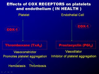
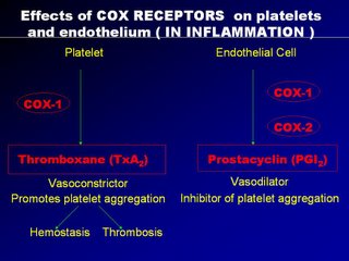
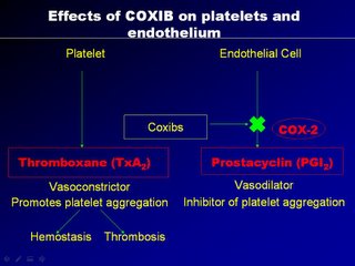
Much has been said recently about the effects of COX2 inhibitors on the heart, in particular coronary artery disease. COX2 is the abbrevation for selective cyclo-oxygenase2 inhibitors, the receptors that help to lessen pain. Examples are drugs like VIOXX, CELEBREX, ARCOXIA etc.

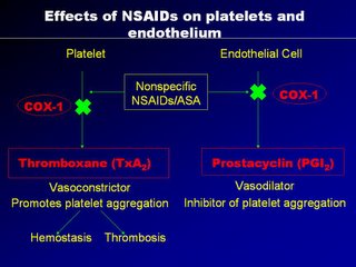
Before the advent of COX2 inhibitors, we had non-selective COX inhibitors (sometimes called COX 1 inhibitors) like NSAIDs (voltaren, cataflam, mobic, aspirin, naproxen, etc.) The problems with COX 1 inhibitors is that, besides alleviating pain, they also cause gastric problems as there are COX 1 receptors in the stomach, and blocking them make them prone to gastric epithelial denudation and the accompanying gastric upset and perhaps gastric bleeding too. In an attempt to lessen the gastric side-effects, COX2 inhibitors were invented.

We became interested in this problem back in 2002 (before the Vioxx controversy exploded on the front page) when the Novartis asked me to look into the problem of "COX2 and Atherothrombosis". Their interest may have been prompted by a paper on CLASS study, that was published in BMJ in June 2002.

Having looked into the issue, I agreed with Novartis that COX2 is potentially harmful for patients suffering from CAD. There were obvious pathogenetic mechanisms to explain this harm. When I examined the CLASS study and also the VIGOR study, I was very surprised that the FDA approved the COX2 as there were inconsistencies in the data pesented. When I mentioned these in the talks that I subsequently gave on behalf of Novartis entitled "COX2 and atherothrombosis", the audience (remember, this was in yr 2002) were quite sceptical.

You may wish to know that the VIGOR study compared the effects on the stomach of ROFECOXIB (Vioxx) against NAPROSYN. CVS risk was a post-hoc analysis which showed an increase incidence of major adverse cardiac events with the use of rofecoxib. The investigators concluded that this could be due to the cardio-protective effects of naprosyn and the FDA did not pursue the matter.

In the CLASS study, the investigators compared the gastro-toxic effects of Celecoxib against a Ibuprofen and Diclofenac. Again, it was not a CVS effects study, and the CVS effects was very much a post-hoc subgroup analysis. In this study, there was no increased incidence of major adverse cardiac events in the celecoxib group. This may be partly because many of the subjects on celecoxib, were also taking aspirin as prevention against CAD. One way of avoiding major adverse cardiac events with the use of COX2 inhibitors is to use it with a small dose of aspirin.

The Theory on COX2 and the heart

Firstly, it is important to note that many tissues in the body has cells that expresses the COX receptors. These tissue include gastric mucosa, fibrous tissues in the joints, and more importantly, platelets and endothelial cells of the artery wall. These tissue have many kinds of COX receptors. Of importance are the COX1 receptors (which are non-specific) and the COX2 receptors which are specific and selective.

These receptors are stimulated by pain and inflammation. Generally speaking, stimulation of the COX receptors produces pain and inhibition of these receptors reduces pain. Although these actions are important in joint pains and inflammation, other effects are more important as far as artherothrombosis is concern.

Almost as if nature must maintain everything in balance, when the COX1 receptors in the platelets are stimulated, it produces thromboxane A2 which causes platelet aggregation and vasoconstriction. When the COX1 receptors in the endothelial cells are stimulated, it produces prostacyclin which causes vasodilatation and also inhibit platelet aggregation. The crucial factor is that the nucleated endothelial cells has COX2 receptors whereas the non-nucleated platelets do not have COX2 receptors. Therefore during episodes of inflammation, platelets aggregate and vasoconstriction is triggered. But those actions are opposed by the prostacyclin from the endothelial cells with counteraction of platelet non-aggregation and vasodilation.

A "yin-yang" like hemostatic balance is maintained and the artery is largely un-affected. Now when we use NSAIDs as anti-inflammatory agents, NSAIDs block the non-selective COX1 receptors equally on the platelets and endothelial cells, and so the hemostasis is balance. But with the use of COX2 agents, they can block COX1 receptors in platelets and endothelial cells (thats okay) but they block selective COX2 receptors, located only in the endothelial cells, thereby diminishing the prostacyclin, so vital for vaso-dilatation and platelet non-aggregation.

The following four slides may somewhat provide a better picture of what's happening (please email me for a copy of the slides).


The result is unopposed vasoconstriction and platelet aggregation. These effects are potentially deletarious in anyone with CAD (known or silent). It's good to note that osteoarthritis affects mainly the elderly, a population at high risk of CAD. Basically, COX2 inhibition is theorectically pro-atherogenic. These was elegently demonstrated in a study by scientist Dr Herman in an study published in Circ. Vol 104 page 820, yr 2001 entitled "Effects of selective cyclooxygenase 2 inhibition on vascular responses and thrombosis in canine coronary arteries".

What was most upsetting is that FDA probably knew about it and did nothing about it. And I quote from the editorial on the CLASS study published in BMJ 1st June 2001.

"Publishing and distributing overoptimistic short term data using post hoc changes to the protocol, while omitting disappointing long term data of two trials, which involved large numbers of volunteers, is misleading. While some of the problems related to CLASS were partially covered in the news sections of BMJ 11 and other journals, it was not emphasised how flawed the trial actually was, and how inadequate the authors' justifications. Consequently, CLASS may still be relied on by many physicians without reference to these flaws. In our experience most still believe the findings published originally. For example, most of 58 physicians attending an osteoarthritis workshop in Berne, Switzerland, in December 2001 had not realised that CLASS was seriously biased."

I therefore believe that on this issue, vioxx has a case to answer.

Signs of a heart attack

This is part eleven of the series the heart of the matter. Part ten can be found here and the disclaimer can be found here.

A heart attack usually causes severe chest pains which is usually over the center of the chest, but feels deep seated. The pain sometimes radiates to the throat, jaw or inner aspect of the left arm. It is usually unprovoked or can also occur following exertion.

The pain, sometimes described as heaviness, crushing, tightness, or shortness of breathe, is usually so severe that it is accompanied by profuse cold sweat, nausea and vomiting. Often, in old people, the pains can be very atypical. Heart attacks in the elderly, can present as faints, loss of consciousness, nausea, vomiting, or just feeling generally unwell. Often, heart attacks can occur silently, especially in diabetics, and only to be discovered on incidental ECG recording for other reasons.

It is fair to say that in patients of the coronary risk group (two or more coronary risk factors), any chest pains should be checked out, the more unwell, the sooner. Better to be overcautious and safe, then to be late and sorry. The ECG is a simple non-invasive means to diagnose a heart attack.


Your doctor may see an ECG such as the one above where the ST elevation is indicative of a heart attack.

Besides an ECG, certain blood test can also be used to diagnose a heart attack. The blood test are to estimate the level of cardiac enzymes in the blood. When heart muscles die, they release enzymes (certain cellular proteins that help in cellular functions). The higher the level, the greater the degree of heart muscle damage.

Some of the heart uscle enzymes that we routinely measure, are SGOT, SGPT, LDH, CPK, CKMB, Troponin T, Troponin I, and others. Some of these blood test can be done by the bedside, eg Troponin T, so that the results can be known immediate, and a definite diagnosis made. REMEMBER TIME IS HEART MUSCLE. For a definite diagnosis of heart attack WHO (World Health Organization) recommends at least two of the three criteria being present. The criteria are definite history suggestive of heart attack, ECG changes of heart attack, or blood test suggestive of a heart attack.

Next we discuss what to do in case of a heart attack.

What happens in a heart attack?

This is part ten in the series the heart of the matter. Part nine can be found here and the disclaimer can be found here.

Coronary artery disease is the building up of cholesterol in the wall of arteries of the heart. This build up occurs insidiously. When it reaches a significant degree, it may cause chest pains on effort, relieved by rest (stable angina). This gradual development occurs in the minority of patients. Usually, the slowly progressive cholesterol plaque breaks causing small blood clots in the artery wall to form. If the blood clots are smaller then the vessel size, the clot flows downstream and gets lodged in a small vessel without any consequence to the patients. He may not even know it. However, if the plaque rupture causes a large enough blood clot to form, the size of the blood clot may occlude the whole artery lumen.


Then there is total obstruction to blood flow down that artery, resulting is no nutrition and oxygen to the muscle supplied by that artery. When those tissues are deprived from life-giving oxygen for more then 40 minutes, the cells begin to die, and get rotten. This, results in the chest pains that the patient experience, the ECG changes and also the blood test abnormalities. This then constitutes what is commonly known as a heart attack.

It is important to emphasize that once the artery is occluded, with each passing minute, more muscle cells die and are lost. If blood flow is restored soon, the cells may recover. However, if there is delay in restoring blood flow, the cell death becomes irreversible and the muscle loss becomes permanent. There is then a saying among us medics, "time is heart muscle". If you want us to help save your heart muscle following a heart attack, come as soon as possible. Remember, TIME IS MUSCLE.

In the next part we examine what the signs of a heart attack are.